STORY #1
To regain Visual Recognition
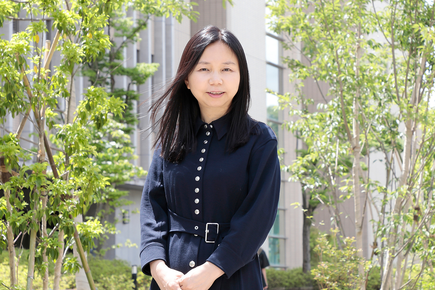
Chieko Koike, Ph.D.
Professor, College of Pharmaceutical Sciences
Creating a Visual Model through International Collaboration
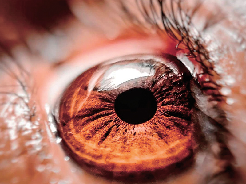
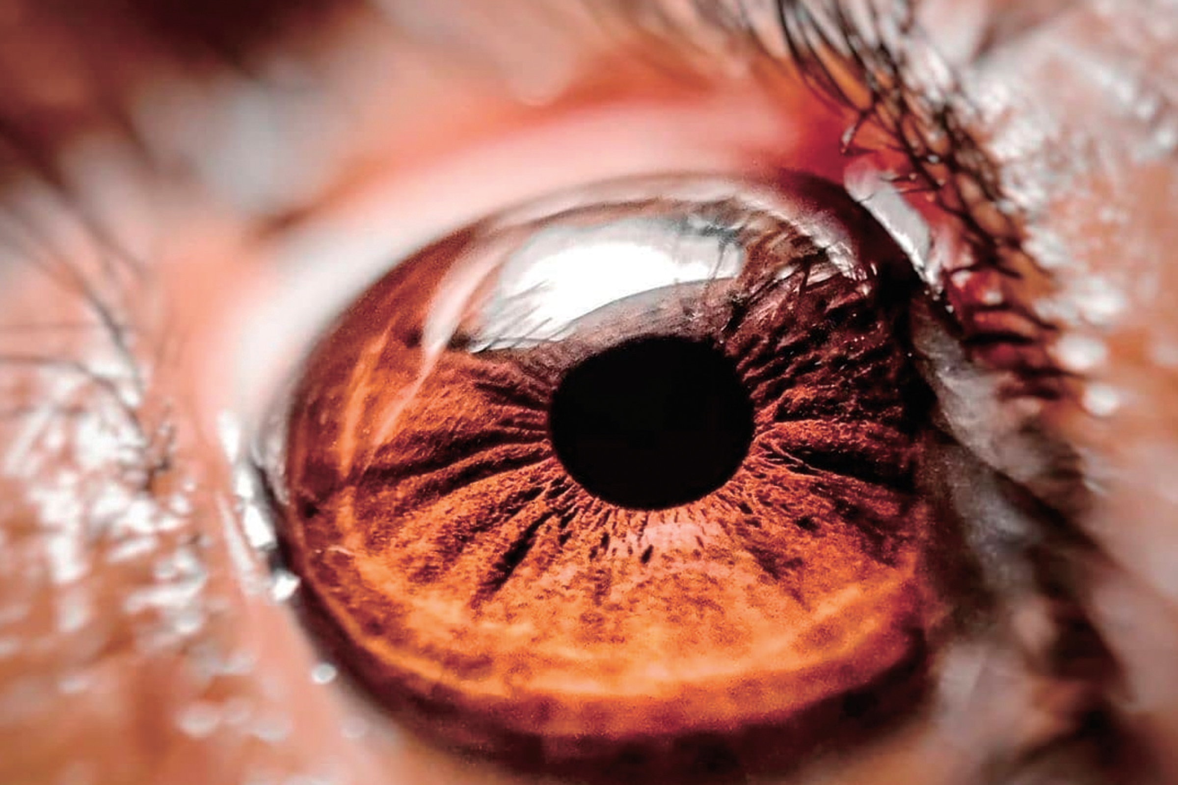
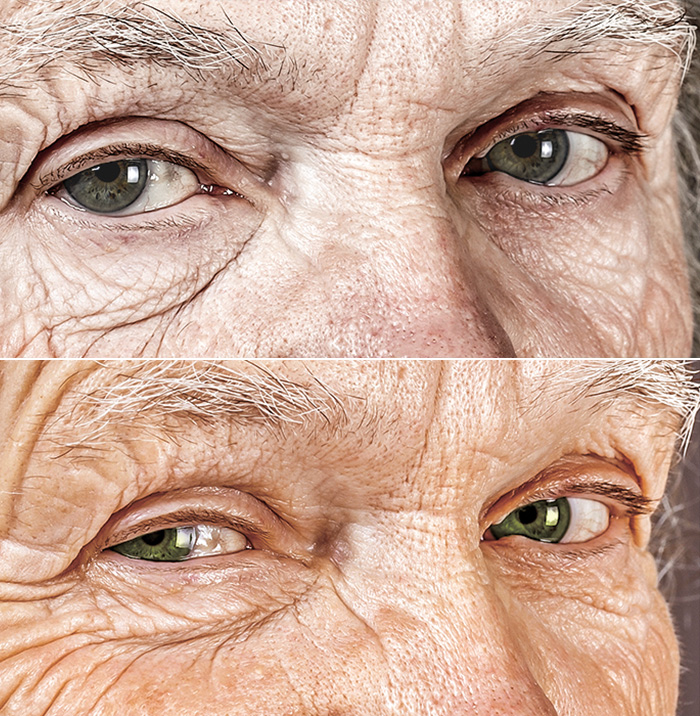
As the society ages, the number of patients suffering from eye diseases is increasing. Although eye diseases rarely lead directly to death, vision loss reduces the quality of life of the patients, causing a loss of employment opportunities for those who want to work and a rise in medical costs. Therefore, it is very important to understand the mechanism of the retina, which is responsible for sensing visual information and transmitting it to the brain, in order to regenerate the visual function.
In 2015, Ritsumeikan University established the Center for Systems Vision Science (CSVS), Japan’s first vision research center for basic science. Chieko Koike, director of the center and a professor at Ritsumeikan University’s College of Pharmaceutical Sciences, explains, “the center has researchers from six departments at Ritsumeikan University, including mechanical engineering, information science, and psychology, as well as life sciences. One of our major strengths is our capability to elucidate the dynamics of the visual system and model it on a computer, and simulations using this model are beginning to be used in clinical settings.”

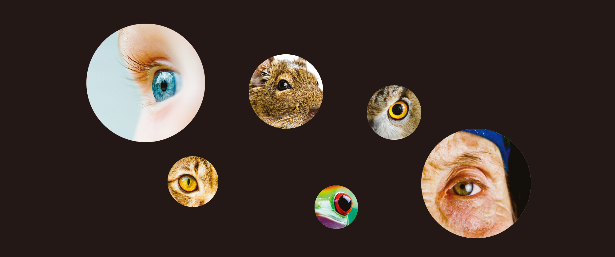
Among the five human senses, vision is the most critical for our quality of life. Information from the sensory organs is typically sent directly to the brain through the sensory nerves. An exception to this is vision. Visual information is processed in the retina before being transferred to the brain. The vertebrate retina develops as an out-pouching from the diencephalon, a part of the central nervous system. Because of its accessibility, the retina has historically been used to study the processing by central neural circuits.
To be more precise, the retina, unlike the nervous systems of other sensory organs, is composed of a number of cells that make up a neural circuit. Photoreceptor cells convert light stimuli into neural signals, which are then sent to bipolar cells, one of the interneurons. At the end of the interneurons are the output cells, the ganglion cells, whose axonal processes enter the optic nerve that leads to the brain. If the photoreceptors signal information and the output cells deliver it to the brain, what is the role of the interneurons that mediate between the input and output? According to Koike, these interneurons are the key to the visual function of the retina.
Koike explains that all retinal photoreceptors respond in a similar way during exposure to light. “There are multiple types of bipolar interneurons, each with different responsiveness to features of the photoreceptor response,” She explains. Roughly speaking, there are two main types of bipolar cells: ON and OFF. The ON bipolar cells are excited when photoreceptors respond to an increase in illumination, whereas the OFF bipolar cells are excited when photoreceptors respond to a decrease in illumination. The two main types of bipolar cells ensure that the signaling of light and dark regions in the visual world is balanced, allowing us to obtain very precise visual information under both conditions. We know that our vision does not see the world as it really is, but rather transforms it to suit our needs. In the case of humans, the most important visual information in our daily lives is the shape of objects. In order to capture the contours of objects, our retina is designed to detect and enhance subtle differences in brightness and darkness. This coordination is made possible by the ON and OFF visual pathways that are created by bipolar cells in the retina. “In our original research, we identified a channel protein called TRPM1 that plays an essential role in the processing of visual information by the ON bipolar cells,” she states. The loss of functional TRPM1 channels results in a disease called congenital stationary night blindness.

Today, Koike is an active leader of the center, which specializes in industry-academia-government collaboration and international cooperation, and she herself presides over international joint research projects. However, she remembers that her international collaboration was kickstarted when she encountered a problem with publishing her research on the channel protein. When Koike began her work over 15 years ago, the identity of the channel responsible for the ON response was one of the most critical gaps in our understanding of retinal function. “We identified the TRPM1 channel as the visual transmission channel of ON bipolar cells and submitted an article on it to Nature, but it was rejected,” Koike recalls. “At first, I didn’t understand why it was rejected, but then I met Professor Steven H. DeVries of Northwestern University, a leading retinal researcher and my current research partner, at a neuroscience research conference in the United States. A Nature paper is meant to report new discoveries, and if the discovery has been published earlier by another group, it loses its value.” Koike had presented their own data at a conference in Japan and then submitted the abstract in English. Through DeVries, she came to understand that competing research groups had learned about her discovery, and that one group added her results to a paper that was already accepted for publication without referring to her abstract. In the world of research, researchers working in the same field are not only rivals but also comrades who belong to the same community. Since Koike had not actively interacted with overseas researchers, her reputation was not known to the reviewers of her paper, which put her at a disadvantage in the review process. Later, when she attended the research conference in the United States, the group that added her result was giving an oral presentation, and Professor DeVries objected to it, saying that the ON bipolar cell TRPM1 channel had first been discovered by a group that included Koike. Owing to this objection, Koike was able to give a short presentation and finally regained her priority.
Since Professor DeVries is a renowned researcher, Koike had wanted him to come to Japan to give a lecture on his research; however, the resources to host him were not initially available. In the meantime, a joint research project with several researchers in Japan, funded by a research grant from Ritsumeikan University, led to the establishment of a research center on the retina, namely the Integrated Vision Research Center, IVRC, the precursor of CSVS. This made it possible for Koike to invite Professor DeVries to speak at a symposium to commemorate the establishment of the IRVC, and their reunion led to their joint research.
“There are a number of advantages to collaborating with Professor DeVries,” Koike explains. “He works with OFF bipolar neurons which is complementary to my own research on ON neurons. Just as some of the techniques that we have developed to study ON bipolar cells are useful for his study of OFF bipolar cells, some of the techniques that he has developed for OFF bipolar cells can help us to study the ON type.” Professor DeVries also enjoys his visits to Ritsumeikan University. When he was in training, both of his mentors had been invited to attend the Taniguchi Symposium, which was held on the shore of Lake Biwa, and they fondly recalled the experience.
Another feature of Professor DeVries’ team is that they use ground squirrels instead of mice as their experimental animals. Koike’s goal is ultimately to learn more about human vision, but the design of both human and non-human primate retinas differs in important ways from that of other mammals, and thus mice, which are widely used as experimental animals, are not suitable for reproducing all aspects of human vision. Looking back at the evolution of vision, vertebrates originally had four color types of vision: red, green, blue, and ultraviolet. Apart from mammals, most living vertebrates retain four-color vision. However, mammals became nocturnal in the Mesozoic Era to escape from dinosaurs and lost two color channels, green and blue, becoming dichromatic. Old World primates, including humans, reacquired a third color channel through a gene duplication that enabled them to see red and green using a mechanism that had originally allowed them to see only red. Furthermore, due to the structure of our skull, both eyes are deeply embedded in the sockets of the eye, and our field of vision always extends to the front of our face. Since the same point on the retina is consistently at the center of the visual field, the color-sensing cells are concentrated in that area, the fovea, which is also adapted for high-acuity vision. The color-sensing cells comprise only 5% of the entire retina, and the rest of the cells can only detect light and dark. In other words, the human retina is heterogeneous between the center of the visual field and the rest of the retina. The mouse’s vision is dichromatic, and its retina is relatively homogeneous, so it cannot reproduce our color perception. On the other hand, ground squirrels are diurnal, and are one of the few mammals that have more color-sensing cells than light- and dark-sensing cells. In the joint research conducted by Koike and Professor DeVries’ team, researchers are comparing the mechanism of information processing in squirrels and mice and creating iPS cells from ground squirrel cells to create a three-dimensional retina.
Koike has had many other encounters with experts and is now able to collaborate not only with university researchers but also with professionals in the clinical medicine field. When it comes to retinal regeneration, the mechanism of the retina is also relevant to clinical professionals, and they are interested in exploring the treatment policy of patients using simulations. CSVS is now the core of international research and has become a center for a wide range of research, including the mechanism of retinal development in evolution, elucidation of retinal circuits, and mathematical simulation of vision.
The five senses, including vision, are used simultaneously and in coordination. Even though the researchers cannot completely regenerate the retina for now, if they can understand the relationship between the vision and the other senses, they may be able to combine multiple senses to obtain a visual recognition that will not cause problems in patients’ daily lives. This idea motivated CSVS to also conduct research on how vision is integrated with other senses and how lost visual recognition is regained. As the center director, Koike sets her goal to have CSVS continue to contribute to clinical practice as Japan’s basic vision research center to improve the QoL of people with eye diseases and visual impairment.
- Chieko Koike, Ph.D.
- Professor, College of Pharmaceutical Sciences
- Specialties: molecular and cellular biology, pathology, electrophysiology, behavioral biology.
- Research Theme: Understanding the network dynamics of the vertebrate retina and its development.